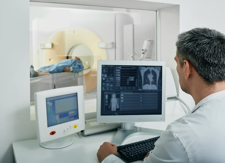
A chest CT is a common imaging study that offers your healthcare provider a close and detailed look inside your chest.
If your healthcare provider has recommended you get a chest CT scan, then you probably have some questions about what to expect from the scan, and how to prepare for your chest CT.
Let’s dive into what a chest CT scan is, how it’s different from other imaging techniques, and what you should know about its safety and radiation concerns.
We’ll help you understand why your doctor may have recommended a chest CT scan.
What is a chest CT scan?
A chest CT scan is a sophisticated medical imaging scan that allows doctors to see detailed pictures of your chest’s interior.
Unlike standard photographs, which capture images from the outside, a chest CT scan uses special equipment to create cross-sectional images (also called slices).
This scan provides your healthcare provider with a comprehensive view of your chest, including your lungs, heart, and the surrounding area, giving them invaluable insights into your health.
How does a chest CT scan differ from other imaging studies?
A chest CT scan is known for its ability to provide exceptionally detailed images of the internal structures within your chest.
This level of detail is important when diagnosing conditions that might not be as visible with less detailed methods.
Moreover, the speed at which a chest CT scan can be performed is a significant advantage, offering quick and accurate diagnoses that are essential in managing health efficiently.
It’s this combination of speed and detail that often makes chest CT scans a preferred choice for exploring complex issues within the chest.
What should I know about the safety and radiation concerns in chest CT scans?
Chest CT scans do involve exposure to a small amount of radiation, but it’s important to balance this with the benefits of obtaining a detailed diagnosis.
Advances in technology have significantly reduced the amount of radiation patients are exposed to during a CT scan.
Additionally, healthcare providers and technicians are trained to use the lowest possible radiation dose that still provides clear images for an accurate diagnosis.
If you have concerns, discussing them with your healthcare provider can provide reassurance and help weigh the benefits against the risks in your specific situation.
Common conditions diagnosed with chest CT scans
A chest CT can be a vital step in diagnosing a variety of conditions, providing detailed images that help pinpoint the cause of your symptoms, and helping to outline a treatment plan.
We’ll explore some of the common conditions that chest CT scans can help diagnose, from lung diseases to heart-related issues.
Which symptoms or signs might prompt a doctor to recommend a chest CT scan?
A wide range of symptoms can lead a doctor to recommend a chest CT scan. Persistent cough, difficulty breathing, chest pain, and unexplained weight loss are just a few symptoms that might prompt further investigation.
These symptoms can be signs of underlying conditions that require detailed imaging to diagnose accurately.
A chest CT scan offers a way to look closely at the structures of your chest, including the lungs and airways, to identify any abnormalities that might explain your symptoms.
This diagnostic tool is particularly useful for evaluating complex cases where other methods might not provide sufficient detail.
How does a chest CT scan assist in diagnosing lung diseases like pneumonia or cancer?
Lung diseases, including pneumonia and cancer, can significantly affect your health, and early diagnosis is key to effective treatment.
A chest CT scan plays a critical role in diagnosing these conditions by providing detailed images that show the lungs and surrounding tissues in great detail.
This level of detail is invaluable for developing an effective treatment plan tailored to your specific condition.
Can a chest CT scan detect heart-related issues or vascular abnormalities?
A chest CT scan can be instrumental in detecting heart-related issues and vascular abnormalities.
Although we often associate chest CT scans with lung diagnostics, they are equally capable of providing insights into the heart’s structure and the major blood vessels that run through the chest.
Conditions such as aortic aneurysm (bulges in the main artery leaving the heart) and other vascular issues can be clearly seen on a chest CT scan. This imaging technique can also assess the extent of these conditions, offering crucial information.
What to expect during a chest CT scan
Knowing what happens during a chest CT scan can help ease your nerves before the scan, and help you to prepare.
We’ll walk you through what happens before your scan, as well as during a chest CT, to make it as straightforward and comfortable as possible.
What do I need to do before my chest CT scan?
Before your chest CT scan, you will receive specific instructions from your healthcare provider.
Generally, these can include guidelines about eating or drinking. For some scans, you may be asked to avoid eating for a few hours beforehand.
You’ll also be advised to wear comfortable, loose-fitting clothing and to remove any jewelry or metal objects that could interfere with the imaging.
In some cases, you might be given a contrast material to drink, or you may receive it as an injection. This contrast helps highlight certain areas in your chest, making the images clearer and more detailed.
Your healthcare provider will let you know exactly what you need to do to prepare, so don’t hesitate to ask questions if anything is unclear.
What happens during a chest CT scan?
You’ll be asked to lie down on a motorized table, which slides into the center of the CT scanner, which is a large, doughnut-shaped machine.
Once you’re positioned correctly, the technologist will step into an adjacent room but can see, hear, and speak with you at all times through an intercom.
As the scan begins, the table will move slowly through the scanner, which rotates around you, capturing images from different angles.
You may be asked to hold your breath for a few seconds to ensure clear images are captured without the movement caused by breathing.
Is the process of a chest CT scan painful or uncomfortable?
The process of a chest CT scan is generally painless. You might feel a bit of discomfort from lying still on the table or, if a contrast material is used, a warm, flushing sensation when it’s administered.
Some people find the enclosed space of the scanner intimidating, but the actual scanning area is open at both ends, and you’ll be able to communicate with the technologist throughout the scan.
If you’re concerned about feeling claustrophobic or uncomfortable, let your healthcare provider know beforehand so they can help make your experience as comfortable as possible.
How long does a typical chest CT scan take?
A typical chest CT scan is quite quick, often completed within a few minutes once the actual scanning begins.
The total time from when you enter the scanning room until you leave can vary but usually doesn’t exceed 30 minutes. This includes the time needed to prepare for the scan, position you on the table, and perform the scan itself.
You can usually resume your normal activities immediately afterward.
Understanding your chest CT scan results
After a chest CT scan, you might feel a little anxious as you wait for your results, which is very common. It’s important for you to understand what your CT results mean for your healthcare.
Let’s explore the insights your healthcare provider can gain from your scan, who will discuss these findings with you, and the potential next steps in your care.
What will my healthcare provider learn from my chest CT scan results?
From your CT results, your healthcare provider can identify a wide range of conditions, from infections and inflammations to tumors and blood clots.
The scan can reveal the size, shape, and position of any abnormalities, helping to diagnose issues such as lung diseases, heart problems, and even injuries to the chest area.
These insights are essential for determining the best course of action, whether it’s further testing, treatment, or monitoring your condition over time.
Who will explain the results of my chest CT scan?
The results of your chest CT scan will be analyzed by a board-certified, subspecialized radiologist.
After reviewing your scan, the radiologist will compile a report detailing their findings. This report is then sent to the healthcare provider who recommended the scan.
Your healthcare provider, who is familiar with your medical history and current health concerns, will discuss the results with you.
They will explain what the findings mean in the context of your overall health and answer any questions you might have.
What are the potential next steps after receiving my chest CT scan results?
If your scan results are normal or show no cause for immediate concern, your healthcare provider might simply continue to keep an eye on your symptoms or suggest lifestyle adjustments to improve your overall health.
Alternatively, if the scan identifies a specific condition, your healthcare provider might recommend further testing to gather more information or start discussing treatment options, which could include medication, therapy, or in some cases, surgery.
If the scan shows something that requires closer observation, you might be scheduled for follow-up scans to monitor any changes over time.
How to schedule an appointment with us
Our goal is to offer you and your healthcare provider the most informative results possible, and we make it easy for you to get an appointment.
With numerous locations across South Jersey, you’ll find us conveniently located near major highways and key bridges in the region.
We’ll ensure the entire scheduling process is as effortless as possible for you. Above all, we are here to help you.
Reach out to us at any of the following locations to book an appointment:
- Haddonfield Office – Haddonfield, NJ
- Marlton (Greentree) Office – Marlton, NJ
- Medford Office – Medford, NJ
- Mount Laurel Office – Mount Laurel, NJ
- Moorestown Office – Moorestown, NJ
- Route 73 (Voorhees) Office – Voorhees Township, NJ
- Sewell (Washington Township) Office – Sewell, NJ
- Turnersville Office – Turnersville, NJ
- Voorhees (Carnie Boulevard) – Voorhees Township, NJ
- West Deptford Office – West Deptford, NJ
- Willingboro Office – Willingboro, NJ
Learn more about the board-certified, subspecialized radiologists who read, analyze and interpret the findings here at South Jersey Radiology Associates.
Frequently Asked Questions
A chest CT scan is a detailed imaging technique that uses X-rays to create comprehensive pictures of your chest and lungs.
Unlike X-rays, a chest CT scan provides more detailed images, and unlike MRI, it uses X-rays instead of magnetic fields to capture these images.
Yes, while chest CT scans involve radiation, the benefits often outweigh the risks, and precautions are taken to minimize exposure.
Symptoms such as persistent cough, difficulty breathing, chest pain, or fever could lead a doctor to recommend a chest CT scan.
It provides detailed images that can reveal abnormalities, infections, tumors, or lung diseases like pneumonia.
Yes, it can identify heart-related issues and vascular abnormalities by providing clear images of the heart and blood vessels.
You may need to avoid eating or drinking for a few hours and remove metal objects that could interfere with the scan.
During the scan, you’ll lie on a table that slides into the CT scanner, and it’s usually quick and painless.
No, the process is not painful, though some may find lying still in the scanner uncomfortable.
A chest CT scan typically takes between 10 to 30 minutes, depending on the specifics of the scan.
Your healthcare provider will gain detailed insights into the condition of your lungs, chest wall, heart, and major vessels.
A radiologist will analyze the images, and your healthcare provider will explain the results to you.
Depending on the results, next steps may include further testing, treatment plans, or monitoring of the condition.


