Can you imagine not being able to breathe or catch your breath? More than 14 million Americans don’t have to imagine it, because they suffer from chronic obstructive pulmonary disease (COPD), a progressive lung disease that is a leading cause of death in the United States.
Smoking, a major cause of COPD, kills more American women each year than breast cancer. In the United States, COPD is especially prevalent in women and people over 65.
Medical technology, however, has made it easier to monitor and assist in the treatment of COPD through the use of chest CT (Computed Tomography) scans. So, if you have been diagnosed with COPD, your healthcare provider will probably recommend regular chest CT scans.
COPD and Its Impact on the Lungs
Chronic Obstructive Pulmonary Disease can make it extremely difficult to breathe by restricting the flow of air into and out of the lungs. Let’s find out more about this disease and its impact on the people it affects.
What is COPD and How Does It Affect The Lungs?
With COPD, less air flows in and out of your airways. The airways in your lungs become more narrow due to being swollen and thickened. This leads to the destruction of the walls between the air sacs in your lungs and those sacs inside the airways lose their ability to stretch and shrink back.
How Does COPD Impact Breathing and Lung Capacity?
In COPD, the airways become inflamed and narrowed, making it difficult to breathe. The air sacs in the lungs, called alveoli, can become overinflated due to trapped air. This hyperinflation reduces the efficiency of gas exchange in the lungs, making it harder for oxygen to enter the bloodstream and carbon dioxide to be removed.
The chronic inflammation seen in COPD can damage the lung tissues over time, leading to scarring and fibrosis. This further reduces lung elasticity and function, contributing to breathing difficulties.
COPD can also cause increased mucus production in the airways, leading to congestion and further narrowing of the already compromised air passages. This can make breathing even more challenging.
How Can COPD Progress Over Time?
As COPD progresses over time, it may become more difficult to breathe, and you may experience more symptoms more often, as well as flare-ups, and other complications. Some of these may become so serious that emergency medical care could be necessary.
What a Chest CT Scan Can Reveal About Your COPD
A chest CT scan can help diagnose COPD, and show the type of COPD (like emphysema or chronic bronchitis) and its progression and severity. A chest CT scan can also help your healthcare provider if surgery is necessary.
How Does A Chest CT Scan Work?
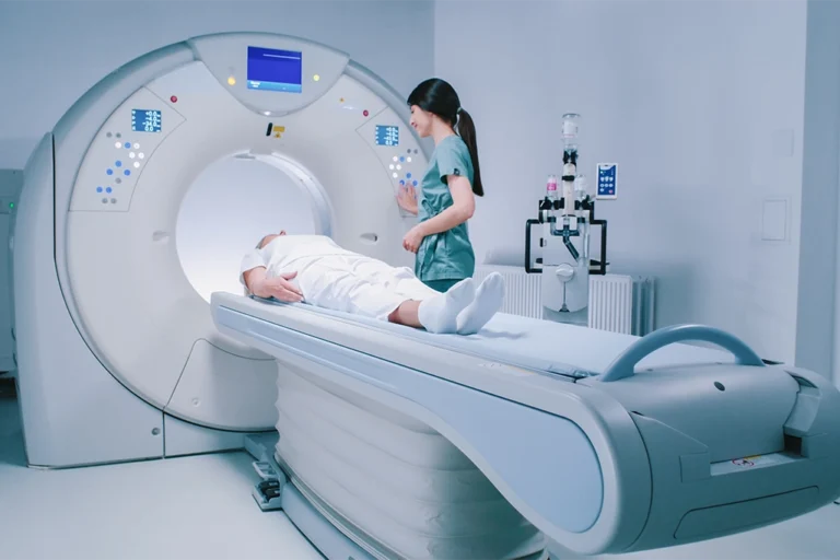
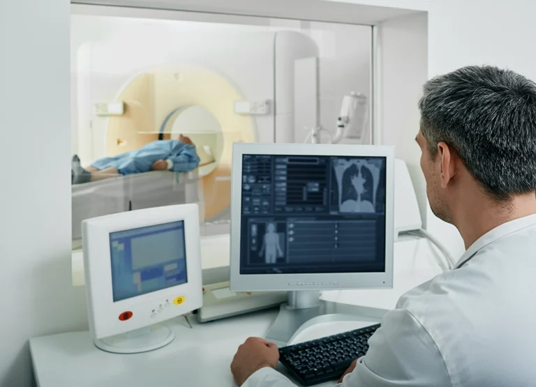
In a chest CT scan, an X-ray beam moves in a circle around your body and chest. It takes many images (called slices) of the lungs and inside the chest. A computer processes these images and displays it on a monitor. The technologist will ask you to breathe in, breath out, or hold your breath to get good quality images of the structures in your chest. The scan usually takes less than 30 minutes.
View our available chest CT appointments at a location near you today
What Are The Specific Findings A Chest CT Scan Can Reveal in COPD Patients?
A chest CT scan can reveal many things about Chronic Obstructive Pulmonary Disease patients, including identification of the presence of any infections or embolisms, and whether or not emphysema is present. The scan can show thickened airway walls, a sign of COPD, which is caused by chronic inflammation and it can also show mucus blockages in smaller airways, which can indicate chronic bronchitis, another aspect of COPD.
How Does A Chest CT Scan Indicate the Severity of COPD?
A chest CT scan can help indicate the severity of COPD by examining the extent of lung damage, emphysema, bronchiectasis, bronchial wall thickening, and pulmonary hypertension. A chest CT scan can show changes in emphysema, air trapping, and chronic bronchitis, which are all characteristic features of COPD. Additionally, the presence of pulmonary hypertension can suggest advanced stages of COPD.
Monitoring The Progress of COPD With Chest CT Scans
The advances in modern medical technology, like today’s chest CT imaging, makes for a far better monitoring process for many diseases, including COPD. Let’s find out more about this monitoring process and how it helps people with COPD.
How does a chest CT scan help in detecting changes in lung tissue over time?
A chest CT scan can help detect changes in lung tissue over time by providing high-resolution images that show even minor changes in the lungs’ structure, making it easier to track the progression of the COPD.
What specific lung changes will my healthcare provider monitor over time?
Your healthcare provider will monitor specific lung changes over time, including airway inflammation, any narrowing of your airways, lung function decline, any scarring on the lungs, and changes in the appearance of lung tissue on the chest CT scan images.

How often should I get a chest CT scan to monitor COPD?
The frequency can vary depending on the individual’s condition, medical history, and history of smoking. In general, chest CT scans for COPD may be recommended on an annual or biennial basis to track disease progression and assess how any treatment is going. It is important to follow the guidance and advice of your healthcare provider of how often you should have a chest CT scan.
The benefits of getting regular chest CT scans
Knowing is always a lot less stressful than not knowing. Getting regular chest CT scans can ease your mind, whether or not you’ve been diagnosed with COPD.
How can regular CT scans assist in managing COPD symptoms?
Regular chest CT scans are extremely helpful in managing COPD symptoms because they can track the progress of the disease with a great degree of accuracy. This means your healthcare provider can alter your treatment plan to offer you a better outcome.
What preventative roles do CT scans play in managing COPD-related complications?
There are many health complications that can be related to COPD, like heart disease and other issues and chest CT scans play an important preventive role in helping treat these complications.
How do chest CT scan results help in creating and adjusting plans?
If you have COPD, regular chest CT scans can chart the progress of the disease and evaluate various treatments you are receiving, thus allowing your healthcare provider to develop even more effective treatment plans.
How to schedule an appointment with us
Our goal is to offer you and your healthcare provider the most informative results possible, and we make it easy for you to get an appointment.
With numerous locations across South Jersey, you’ll find us conveniently located near major highways and key bridges in the region.
We’ll ensure the entire scheduling process is as effortless as possible for you. Above all, we are here to help you.
- Haddonfield Office – Haddonfield, NJ
- Marlton (Greentree) Office – Marlton, NJ
- Medford Office – Medford, NJ
- Mount Laurel Office – Mount Laurel, NJ
- Moorestown Office – Moorestown, NJ
- Route 73 (Voorhees) Office – Voorhees Township, NJ
- Sewell (Washington Township) Office – Sewell, NJ
- Turnersville Office – Turnersville, NJ
- Voorhees (Carnie Boulevard) – Voorhees Township, NJ
- West Deptford Office – West Deptford, NJ
- Willingboro Office – Willingboro, NJ
Learn more about the board-certified, subspecialized radiologists who read, analyze and interpret the findings here at South Jersey Radiology Associates.
Frequently Asked Questions
COPD, or chronic obstructive pulmonary disease, is a chronic inflammatory lung disease that obstructs airflow and makes breathing difficult.
COPD reduces lung capacity by damaging the airways and air sacs, leading to shortness of breath, and reduced oxygen intake.
COPD can worsen gradually, causing increased lung damage, more severe breathing difficulties, and a higher risk of complications.
A chest CT scan uses X-rays to create detailed cross-sectional images of the lungs and the chest to detect abnormalities.
A chest CT scan can show emphysema, airway thickening, and other lung abnormalities related to COPD.
The scan can reveal the extent of lung damage, such as the amount of emphysema and bronchial wall thickening, which indicates the severity of COPD.
It provides detailed images that allow healthcare providers to monitor lung tissue changes, and to track disease progression.
The frequency of chest CT scans varies but is generally recommended annually or biannually, depending on the severity and progression of COPD.


