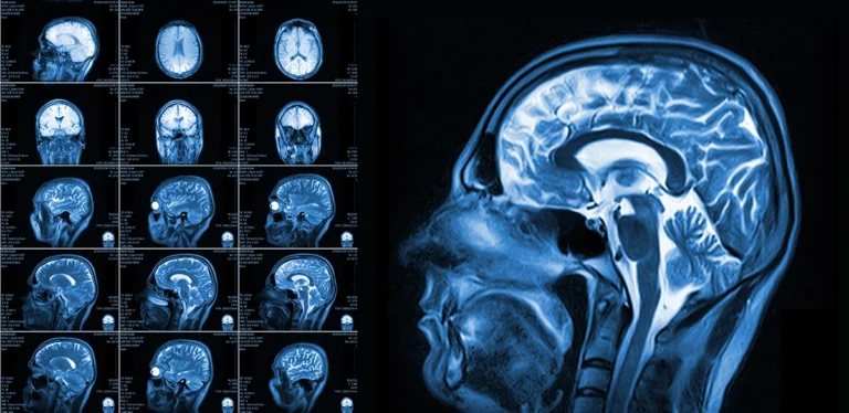
An MRI scan is uniquely suited for assessing a traumatic brain injury (TBI) because it offers detailed, high-resolution images that can detect subtle changes in brain tissue that other scans might miss.
MRI scans are particularly helpful because they can pinpoint areas of inflammation, bleeding, or microscopic damage, providing critical information for accurate diagnosis and effective treatment planning.
We’ll show you why MRI scans are recommended for detailed imaging, why MRIs are preferred over other scans for TBIs, how an MRI can measure the effects of a brain injury, and how MRI scans help detect an injury when your symptoms are unclear.
MRI scans are recommended when more detailed brain imaging is needed
Experiencing a traumatic brain injury is a frightening event. An MRI scan after a traumatic brain injury will give your healthcare providers the needed information to assess the extent of your injury. The detailed scan is often the preferred scan for this type of injury, as it shows even subtle injuries that may not be seen on other scans.
When is an MRI necessary for diagnosing a traumatic brain injury?
An MRI might be necessary if your traumatic brain injury is not clearly seen on a CT scan. Conditions that persist after an injury may be subtle, and may not be visible on a CT scan.
An MRI will help by showing even small injuries that can be present after a traumatic brain injury, allowing for accurate diagnosis and treatment.
How do healthcare providers determine whether an MRI is needed after a head injury?
After a head injury it is important to understand the location and extent of damage to the brain. Often a CT scan is the first scan when a brain injury happens, as it is faster than an MRI and can quickly identify urgent injuries needing immediate attention.
However, an MRI can show where subtle damage has occurred, as it has higher resolution and shows more detail than does a CT scan. If your healthcare provider suspects subtle damage that is not seen on a CT scan, they may recommend an MRI.
When is an MRI the preferred scan over other imaging tests?
After a traumatic brain injury there may be damage to soft tissues of your brain that is not easily seen on a CT scan. If symptoms such as headaches or dizziness continue, but injury is not seen on a CT scan, an MRI may provide the needed information to help identify the source of your symptoms.
View our available MRI appointments at a location near you today
An MRI scan is preferred over a CT scan for certain brain injuries
After a traumatic brain injury, both CT scans and MRI scans can play important roles. Brain injury can be both dramatic and subtle, and different types of injuries can appear more clearly on some scans than others. Depending on the nature of your injury, and the symptoms you are experiencing, your healthcare provider may prefer that you have an MRI scan to make sure no subtle injuries are present.
How do MRI and CT scans differ in detecting traumatic brain injuries?
A traumatic brain injury is often an emergency situation. At first, the extent of damage is unknown, and a quick scan to assess immediate interventions is needed.
A CT scan is ideal for this, as it does show injuries that may be life-threatening. These can include skull fractures and significant brain bleeds. A CT scan is also much quicker than an MRI scan, which is important when a situation is urgent.
When more subtle injuries are present, an MRI scan is useful in showing tissue damage or tears in detail that may not be clear on a CT scan.
What types of brain injuries are more visible on an MRI than on a CT scan?
Injuries that require a high level of image resolution, like microbleeds, bruising, and swelling, will benefit from the enhanced imagery of an MRI scan. An MRI scan has more sensitivity and detail than does a CT scan, so injuries that may not be seen at first can be found through an MRI.
When do doctors recommend an MRI after an initial CT scan?
Your healthcare provider might suggest an MRI even after an initial CT scan, especially if they suspect that there may be more subtle injuries that are present. A CT scan is preferred in situations where speed is important, and to treat more immediate and obvious injuries.
Beyond that, an MRI can help identify the source of continued symptoms or suspected injuries that may not be clearly seen on a CT scan. MRI scans can also help to monitor your condition following an injury.
MRI scans can detect the subtle and long-term effects of a TBI
After a traumatic brain injury, there are often both immediate and long-term concerns, and your provider needs precise information about your condition. Sometimes, injuries are not obvious, or they may develop gradually over time. MRI scans are a useful tool to help identify injuries that may not be seen at first.
How does an MRI detect microbleeds and other small brain injuries?
An MRI is particularly effective at detecting microbleeds and small brain injuries because it captures highly detailed images of the brain’s soft tissues using powerful magnets and radio waves. MRI scans can highlight tiny areas of bleeding or subtle tissue damage by showing differences in how blood products interact with magnetic fields.
These imaging techniques reveal microbleeds as distinct dark spots, clearly distinguishing them from surrounding healthy tissue. By identifying even minor injuries, an MRI provides your healthcare provider with critical insights into the extent and location of brain damage, helping guide appropriate treatment and recovery decisions.
How can an MRI scan identify long-term brain damage?
An MRI scan can be helpful in identifying the presence and type of long-term brain damage. The scan can show atrophy (or shrinking) of the brain that can be a long-term effect of a traumatic brain injury.
It will also show lesions or scarring that might be present after an injury. These will appear as dark spots on the scan. Structural damage to the brain can be identified through the magnetic fields that capture even small changes or abnormalities in brain tissue.

How does an MRI help to guide treatment after a TBI?
The detailed images from an MRI scan will show your healthcare provider the location, extent, and nature of brain tissue damage following a traumatic brain injury. If surgery is needed, the images can help surgeons understand what is happening in the brain, and how to best approach surgery.
It will show them if there are blood clots that should be removed, for example, and the size and location of the clot. Knowing what types of injury are present, as well as the location and extent of the injuries, is helpful in guiding treatment after a traumatic brain injury.
An MRI scan detects brain injuries even when symptoms are unclear
After a traumatic brain injury, it can be difficult to know why you might be having symptoms such as headache or nausea. An MRI scan can help rule out damage to parts of the brain, as well as identify where damage has taken place. Knowing what kind of damage has occurred after an injury can be helpful in managing symptoms and planning for future care.
How does an MRI scan help to confirm or rule out a TBI?
The images from an MRI scan can help to show whether your symptoms are related to a traumatic brain injury or to another source. These conditions can cause symptoms that could be similar to those of a TBI.
That way, an MRI scan can identify different types of damage to help chart appropriate treatment, whether for a TBI or other condition.
What types of brain injuries can an MRI reveal that may not be immediately apparent?
After a traumatic brain injury, there may be damage that is not seen right away. An MRI can help identify even small injuries that may cause more harm over time.
These include small strokes, microbleeding, damage to nerves, concussion, or swelling of brain tissue. An accurate diagnosis of these subtle conditions is essential to protect future brain health and plan for the right kind of treatment.
How can an MRI help determine the severity of a traumatic brain injury?
The detailed images of an MRI scan will show the nature, location, and extent of damage to the brain after an injury. The severity of an injury can be assessed through the images from an MRI scan.
These include finding bleeding, swelling, or bruising of the brain. Finding can also show whether the brain has been deprived of oxygen for a time, or whether nerve damage has taken place.
The precise imaging of an MRI scan helps your healthcare provider understand the extent of damage after an injury. That way, they can work to minimize harm and plan future treatments to ease symptoms and aid in your recovery.
How to schedule an appointment with us
Our goal is to offer you and your healthcare provider the most informative results possible, and we make it easy for you to get an appointment.
With numerous locations across South Jersey, you’ll find us conveniently located near major highways and key bridges in the region.
We’ll ensure the entire scheduling process is as effortless as possible for you. Above all, we are here to help you.
Reach out to us at any of the following locations to book an appointment:
- Marlton (Greentree) Office – Marlton, NJ
- Medford Office – Medford, NJ
- Moorestown Office – Moorestown, NJ
- Mount Laurel Office – Mount Laurel, NJ
- Route 73 (Voorhees) Office – Voorhees Township, NJ
- Sewell (Washington Township) Office – Sewell, NJ
- Turnersville Office – Turnersville, NJ
- Voorhees (Carnie Boulevard) Office – Voorhees Township, NJ
- West Deptford Office – West Deptford, NJ
- Willingboro Office -Willingboro, NJ
Learn more about the board-certified, subspecialized radiologists who read, analyze and interpret the findings here at South Jersey Radiology Associates.
Frequently Asked Questions
An MRI is recommended when a more detailed view of the brain is needed, especially if symptoms persist despite a normal CT scan.
Doctors assess the severity of symptoms, neurological function, and initial imaging results to decide if an MRI is necessary for further evaluation.
An MRI is preferred when detecting subtle brain injuries, such as microbleeds or damage to soft tissues, that may not be visible on a CT scan.
CT scans are better for identifying skull fractures and acute bleeding, while MRIs provide a more detailed look at brain tissue damage and smaller injuries.
MRIs can detect microbleeds, diffuse axonal injuries, and subtle changes in brain tissue that are often missed by CT scans.
MRIs use advanced imaging techniques, such as susceptibility-weighted imaging, to reveal tiny blood vessel damage and other microscopic injuries.
By identifying specific areas of brain damage, an MRI helps doctors develop targeted treatment plans and monitor recovery over time.
An MRI provides detailed images of brain structures, allowing doctors to assess the extent of damage and predict potential long-term effects.


