
The human brain is an astonishingly complex organ. It can process information rapidly, store memories, and manage all the functions of our bodies. When someone starts experiencing symptoms that hint at a neurological issue, doctors typically recommend specialized MRI scans to get a clearer picture of what might be happening inside.
A neurologist typically orders a brain MRI, also known as a head MRI, if they suspect an underlying issue, need help diagnosing a condition, or wish to monitor the development or treatment of an injury. Ordering an MRI of the brain does not confirm a diagnosis, but rather signals that your neurologist requires more information to develop the best possible treatment plan. South Jersey Radiology Associates offers advanced MRI services to help you obtain the accurate diagnostic images you need.
What does an MRI of the brain show?
A brain MRI provides detailed images of the brain’s structures, allowing healthcare providers to detect potential abnormalities such as tumors, infections, or brain damage. The results reveal problems concerning the brain’s structure, including tumors, inflammation, and swelling. An MRI can also reveal bleeding, infections, damage from injury, or a stroke, and help healthcare providers determine the causes of headaches or seizures.
Reasons you may need a brain MRI
An MRI scan of the brain can help diagnose issues related to the brain, its nerves, inflammation, inner ear problems, and the spinal cord. Specific diagnoses possible following a head MRI include:
- Blood clot or hemorrhage
- Infection
- Stroke
- Damage associated with epilepsy
- Tumors and cysts
- Causes of headaches and migraines
- Multiple sclerosis (MS)
- Dementia and Alzheimer’s (later stages)
- Hydrocephalus (water on the brain)
- Pituitary gland issues
- Traumatic brain injury
- Issues with brain development or inner ear problems
Imaging’s role in monitoring cognitive function
A brain MRI plays a vital role in the early detection, diagnosis, and monitoring of cognitive conditions. Because it provides detailed MRI images without using ionizing radiation, it is considered a safer option for repeated scans over time, which is essential for monitoring health. For conditions such as Alzheimer’s disease, Amyloid PET scans are typically ordered to check for early signs of the disease, such as plaque buildup and changes in tissue function. In tandem with these scans, MRI exams are crucial for periodically monitoring the brain’s response to therapy, as they can reveal changes in brain size, structure, and function, particularly in areas critical for memory and cognition. This combination of imaging techniques allows for a comprehensive assessment of Alzheimer’s progression and treatment efficacy.
How to prepare for a brain MRI
In the days leading up to your scan, you can typically continue eating, drinking, and taking your medication as usual. However, on the day of the exam, you may be recommended to avoid eating or drinking for the hours leading up to the scan to avoid any interference with activity in your brain. You will be asked to fill out a patient history form prior to your appointment, and will receive specific preparation instructions based on your medical history.
If you are undergoing an MRI with contrast, please plan to arrive around 15 minutes early for your exam.
Items to remove before entering the MRI scanner
When preparing for an MRI scan, remember that it uses a strong magnetic field from a powerful magnet. Before entering the MRI scanner, you must remove any metallic objects that may be on your person, including:
- Hair accessories, such as clips, pins, or bands
- Jewelry, including watches, necklaces, bracelets, earrings, and rings, and body piercings
- Credit cards and debit cards, which can be affected by the magnetic fields
- Hearing aids
- Dentures and any removable dental work that contains metal
- Clothing with metal, avoiding items with buttons, zippers, or metal fasteners (a hospital gown may be provided if necessary)
- Cosmetics, as some makeup products contain tiny metal particles
If you have items like aneurysm clips, or metal plates and staples that can’t be removed, please inform your doctor and our team at the time of scheduling.
View our available brain MRI appointments at a location near you today
What to expect during the procedure
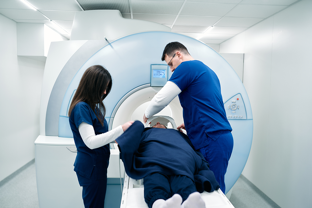
During a brain MRI, you will lie on your back on the MRI bed, which slides into the donut-shaped MRI machine. Once positioned, a plastic head coil is secured around your head; this coil improves the clarity of the MRI images as it interacts with the magnets in the MRI scanner.
How long does a brain MRI take?
Your brain MRI will likely take between 30 and 60 minutes, and it is crucial that you lie completely still throughout this time. The MRI machine generates loud noises; however, earplugs will be provided for your comfort.
You can communicate with the technologist during the procedure using an intercom, and a panic button is located inside the machine in case you feel overwhelmed.
Is a brain MRI painful or uncomfortable?
The MRI procedure itself is not painful. Discomfort is usually related to lying still or feeling claustrophobic.
Our 70 cm wide-bore MRI scanners prioritize comfort by offering up to 20% more space than traditional scanners, catering to those who may feel anxious during imaging scans. It comfortably accommodates patients up to 550 lbs., creating a more relaxed scanning experience.
In addition to our spacious scanners, SJRA offers fast scan MRI technology that can significantly improve your experience. This innovative approach reduces scan times by up to 48%, meaning you’ll spend less time lying still and holding your breath, which helps minimize discomfort and anxiety. The use of advanced AI technology not only speeds up the process but also ensures high-quality images, providing clearer results for your physician. Our softer, lighter GE AIR™ Coils are designed to feel like a comforting blanket and form around your body. With these advancements, we are committed to making your MRI experience as pleasant as possible.
If you are anxious about panicking, discuss options with your doctor. In some cases, you may be eligible to take a mild oral sedative before your appointment. Always inform our team if you plan on taking a sedative beforehand.
What are the side effects of a brain MRI?
There are no side effects from the MRI process itself. However, if your doctor orders an MRI with contrast, there may be potential side effects related to the injection.
Contrast material is a gadolinium-based dye injected into an arm through an IV line. The contrast is used to enhance the quality of the image and check the flow, volume, or supply of blood vessels in different areas. Contrast can be particularly useful in diagnosing MS or monitoring cancer growth.
Side effects associated with the injection or contrast material may include hives, itchiness, nausea or vomiting, and pain at the injection site. If you have kidney problems, please inform your doctor, as this may affect your body’s ability to break down the contrast material.
Understanding and discussing your MRI results
The results of your brain MRI must first be studied and analyzed by a radiologist who has expertise in the area of the body being studied and then shared with your healthcare provider.
Do you get brain MRI results immediately?
No. After your scan is complete, the radiologist sends the scan to your doctor, who will then contact you for a follow-up appointment to discuss the findings and potentially create a treatment plan.
How abnormalities are identified
On a brain MRI scan, abnormalities or lesions appear as dark or light spots that do not resemble normal brain tissue. Healthcare providers look for differences in tissue density, shape, size, and contrast to detect anomalies such as tumors, lesions, hemorrhages, or structural abnormalities.
Brain MRI results and how to prepare
An abnormal result does not necessarily mean there is a serious problem. Approximately 17% of MRI exams conclude with an abnormal result, which simply means the head or brain is not perfectly healthy. Abnormal findings can range from brain lesions and white spots (symptomatic of issues like small strokes, MS, migraines, or B-12 deficiency) to structural damage or inner ear problems.
Signs on a brain MRI that indicate serious conditions like a tumor or a stroke may include:
- Tumor. Irregular masses or lesions, abnormal areas (often highlighted by contrast dye), distortion of brain structures, edema (swelling), and displacement of surrounding tissues.
- Stroke. Areas of restricted blood flow (ischemia), changes in signal intensity in affected regions, and loss of brain tissue volume.
Common benign findings
Research indicates that the most common benign findings on a brain MRI often include:
- Small areas of bleeding (frequently observed in older individuals)
- Areas of increased signal intensity (common in older adults, often related to small blood vessel disease)
- Pineal cysts, arachnoid cysts, and choroid plexus cysts (fluid-filled sacs that are typically asymptomatic)
- Enlarged spaces around blood vessels in the brain
- Sinus mucosal thickening or an enlarged pituitary gland
It is essential to consult a qualified healthcare provider for a final analysis, even if the findings are common or usually benign. Ask questions and seek clear explanations to better understand your health. If any issues are found, your results will help guide a personalized care plan, which may include further tests, specialist consultations, or treatments. Follow-up MRI exams might also be recommended to monitor changes over time.
So, why would a doctor order an MRI of the brain?
At South Jersey Radiology Associates, we are trusted by patients for our expertise and prioritize your health and understanding in all aspects of diagnostic imaging. A brain MRI can be a crucial step in identifying and monitoring neurological issues, providing detailed insights that aid your healthcare provider in making informed decisions about your treatment.
- A brain MRI is essential for diagnosing various neurological conditions, including strokes, tumors, and multiple sclerosis.
- This imaging technique allows for the detection of abnormalities without exposure to ionizing radiation, making it safer for repeated assessments.
- Proper preparation is vital for accurate results, including the removal of metallic objects and following pre-scan instructions.
If you’re experiencing symptoms that may require a brain MRI or if you want to learn more, don’t hesitate to schedule your appointment today by calling us at (888) 909-7572 or scheduling an appointment online today.
Frequently Asked Questions
A head MRI scan can detect various issues related to the brain, nerves of the brain, inflammation in the head, inner ear problems, and the spinal cord. In some cases, an MRI with contrast may be necessary to assess blood flow and improve image quality.
Your doctor may order an MRI scan with contrast if they suspect certain conditions, such as multiple sclerosis (MS), cancer growth, or soft tissue development problems. The contrast dye provides more detailed information in diagnosing these specific conditions. It is important to discuss with your doctor why they have or haven’t recommended an MRI with contrast.
Your doctor may recommend a head MRI scan if you have recently suffered a head injury, experience headaches when sneezing or coughing, confusion, numbness or weakness, muscle weakness or tingling, changes in thinking or behavior, hearing loss, speaking or vision difficulties, pulsating feelings during headaches, headaches in the morning, constant headaches, seizures, vertigo, extreme weakness, or fatigue. However, experiencing these symptoms does not guarantee that something is wrong.
If you are unable to undergo an MRI due to reasons such as claustrophobia or an unremovable metal device in your body, a CT scan can be an alternative. However, a head MRI is generally preferred as it provides clearer and more detailed images. MRIs are safer than CT scans because they use magnets instead of ionizing radiation. It’s important to consult with your doctor about the most suitable imaging option for you.
The cost of a head CT scan without insurance ranges from $270 to $5,000, while a head MRI ranges from $250 to $8,750. Insurance coverage for an MRI scan is likely if it has been recommended by your doctor. Insurance may deny coverage if a cheaper alternative scan, such as a CT scan, could suffice. To save money, consider visiting an independent radiology center, where MRI scans can cost up to 60% less than those at hospital facilities.
Ask about the significance of the findings, potential impact on your health, and next steps.
Depending on the findings, they might recommend additional imaging studies, blood tests, or a biopsy to gather more information.
You can book a head MRI appointment at any of the following locations:
• Marlton (Greentree) Office – Marlton, NJ
• Medford Office – Medford, NJ
• Moorestown Office – Moorestown, NJ
• Mount Laurel Office – Mount Laurel, NJ
• Route 73 (Voorhees) Office – Voorhees Township, NJ
• Sewell (Washington Twp) Office – Sewell, NJ
• Turnersville Office – Turnersville, NJ
• Voorhees (Carnie Blvd) Office – Voorhees Township, NJ
• West Deptford Office – West Deptford, NJ
• Willingboro Office – Willingboro, NJ


