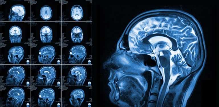
When your healthcare provider recommends a brain MRI to check for signs of a stroke, you may be wondering what it can show, and why it’s important. Even if you’ve had an MRI in the past, this one may feel different because it’s focused specifically on the brain.
A brain MRI can detect the signs of stroke, and can help pinpoint when the stroke may have occurred. It also offers a detailed look at how different parts of the brain may be linked to your symptoms.
If you’re feeling unsure about what this scan is meant to find, you’re not alone. Keep reading to learn how a brain MRI can help guide your care and give your provider the answers they need.
Why your provider recommended a brain MRI scan
When your provider suspects a stroke, a brain MRI is often one of the first ways they look for clear answers. We’ll explain why brain MRIs are especially useful for finding signs of stroke, how they create detailed images of the brain, and what makes them different from other types of scans.
What makes a brain MRI scan a good tool for finding the signs of stroke?
A brain MRI is especially good at picking up early changes in brain tissue, including those caused by reduced blood flow or bleeding. This can make it easier to detect stroke-related damage even in the first few hours after symptoms begin.
MRI scans are sensitive to tiny shifts in water molecules in the brain, which often occur during a stroke. These subtle changes can help your provider see what’s happening in your brain before major damage sets in.
How does a brain MRI create images that can help diagnose stroke?
Brain MRIs use strong magnets and radio waves to create detailed pictures of your brain’s structure. These images can reveal areas that aren’t getting enough blood, as well as spots where bleeding may have occurred.
Different types of MRI settings highlight different brain tissues, making it easier to tell if a stroke has happened, what kind it might be, and how recent it is. This information helps guide decisions about what steps to take next.
What makes an MRI more detailed than other brain scans, like CT?
An MRI can show much finer details in soft tissue than a CT scan, which means it can spot small changes that might be missed otherwise. This can be especially important in the early stages of a stroke or when symptoms are mild.
Because MRIs can capture images from many different angles and settings, they give a clearer picture of the brain’s condition. That level of detail can make a real difference in identifying subtle signs of stroke.
View our available brain MRI appointments at a location near you today
How a brain MRI scan shows the signs of a stroke caused by a blockage
When a stroke is caused by a blockage, it means part of the brain isn’t getting the blood and oxygen it needs. Let’s learn how brain MRIs help spot blockages, what happens to brain tissue when blood flow is disrupted, and how these changes appear in MRI images.
What is a stroke caused by a blockage? How does a brain MRI find it?
A stroke caused by a blockage, also called an ischemic stroke, happens when a clot or other material slows or stops blood from reaching part of the brain. This can quickly lead to damage if oxygen and nutrients are cut off for even a short time.
A brain MRI can detect this type of stroke by picking up changes in the water content of brain tissue where the blockage has reduced blood flow. These changes often appear as bright or dark spots, depending on the MRI settings used.
What happens to brain tissue when blood flow to part of the brain is interrupted?
When blood can’t reach part of the brain, the affected cells begin to break down, and chemical changes take place. This can start within minutes, and the damage spreads if blood flow isn’t restored.
As cells are injured, they release substances that attract water into the area, causing swelling. A brain MRI can capture this swelling and the shift in water balance, giving an early clue that a stroke may be in progress.
How does a brain MRI show areas that may have been affected by a lack of oxygen?
Certain MRI settings are especially sensitive to parts of the brain that have been starved of oxygen. These areas may show up as bright spots on the scan, often before other changes are visible.
This allows your provider to identify affected brain tissue quickly, even if symptoms are still new or unclear. It also helps estimate how recent the stroke might be, which can influence the next steps in your care.
How a brain MRI can show the evidence of a stroke caused by bleeding
When a stroke is caused by bleeding in the brain, it involves different risks and symptoms than a stroke caused by a blockage. This section explains how a brain MRI can detect bleeding, what it looks like on the scan, and how the images help pinpoint the location and size of the affected area.
What is a stroke caused by bleeding of the brain? How can an MRI find it?
A stroke caused by bleeding, also called a hemorrhagic stroke, happens when a blood vessel in the brain breaks and leaks blood into nearby tissue. This can put pressure on the brain and damage the surrounding cells.
A brain MRI can pick up signs of this kind of stroke by detecting blood in places where it doesn’t belong. Different settings on the MRI can help reveal both fresh bleeding and areas where blood has pooled over time.
What does a brain MRI show when there is bleeding inside the brain?
Fresh bleeding often shows up as bright or dark patches on an MRI, depending on the type of scan and how long the bleeding has been present. These patches can appear very clearly against the normal structure of the brain.
As the blood starts to break down, MRI scans can also show changes in color and shape that give clues about how long ago the bleeding started. This helps your provider track how the situation is evolving.
How can MRI images help identify where the bleeding happened and how much area is involved?
MRI scans can map the exact location of the bleeding and show how much of the brain is affected. This is especially important for understanding which parts of the brain might be impacted.
Some areas are more sensitive than others, and seeing the size and position of the bleed helps guide what kind of care is needed. The level of detail an MRI provides makes it easier to spot even small bleeds that might otherwise be missed.

How a brain MRI helps your provider understand your symptoms
When you’re experiencing symptoms that could be linked to a stroke, timing and detail matter. This section explains how a brain MRI can help your provider estimate when a stroke may have happened, connect the results to your specific symptoms, and decide on the most appropriate next steps.
Understanding how your scan fits into the bigger picture can make the process feel more grounded and clear.
How can my MRI results help my provider estimate when a stroke may have happened?
A brain MRI can help estimate how old a stroke might be by showing how brain tissue has changed over time. Different patterns on the scan can suggest whether a stroke is very recent, a few days old, or even older.
Some MRIs can highlight fresh changes in water flow or oxygen levels, while others pick up signs of tissue breakdown or healing. These time-based clues help your provider better match your symptoms to what’s happening inside your brain.
How do my results help my provider to diagnose my specific symptoms?
Your symptoms might include changes in movement, speech, vision, or memory—each linked to different areas of the brain. A brain MRI can help your provider see if there’s damage in a region that lines up with what you’re experiencing.
For example, if you’ve had trouble speaking clearly, the scan might reveal changes in the part of the brain that controls language. This connection helps confirm the cause and gives a clearer picture of what’s happening.
How will my provider use my MRI results to guide the next steps in my treatment?
Once your provider understands what type of stroke may have happened, how recent it is, and which areas of the brain are involved, they can plan the right path forward. The scan helps rule out other causes and gives a clearer view of what kind of care you might need next.
Your MRI results may help decide whether medication, monitoring, or other forms of support are the best fit for your situation. The more precise the information, the better they can match your needs to the right care.
How to schedule an appointment with us
Our goal is to offer you and your healthcare provider the most informative results possible, and we make it easy for you to get an appointment.
With numerous locations across South Jersey, you’ll find us conveniently located near major highways and key bridges in the region.
We’ll ensure the entire scheduling process is as effortless as possible for you. Above all, we are here to help you.
Reach out to us at any of the following locations to book an appointment:
- Marlton (Greentree) Office – Marlton, NJ
- Medford Office – Medford, NJ
- Moorestown Office – Moorestown, NJ
- Mount Laurel Office – Mount Laurel, NJ
- Route 73 (Voorhees) Office – Voorhees Township, NJ
- Sewell (Washington Township) Office – Sewell, NJ
- Turnersville Office – Turnersville, NJ
- Voorhees (Carnie Boulevard) Office – Voorhees Township, NJ
- West Deptford Office – West Deptford, NJ
- Willingboro Office – Willingboro, NJ
Learn more about the board-certified, subspecialized radiologists who read, analyze and interpret the findings here at South Jersey Radiology Associates.
Frequently Asked Questions
A brain MRI offers highly detailed images that help your provider look closely for signs of stroke that other scans may miss.
An MRI can reveal changes in brain tissue where blood flow has been reduced or cut off, helping to locate the affected area.
When part of the brain is deprived of oxygen, the tissue starts to change, and an MRI can capture those changes early on.
Yes, MRI scans can detect bleeding in the brain, and help pinpoint where it happened and how much tissue is affected.
The scan may show areas of abnormal signal where blood has collected or leaked into brain tissue.
Certain changes in the brain appear differently, depending on how much time has passed, which helps estimate when the stroke occurred.
Yes, the scan can connect the location of the brain injury to the functions that may have been affected.
Your provider will use the detailed images to confirm the type of stroke, and decide what treatments or next steps are right for you.