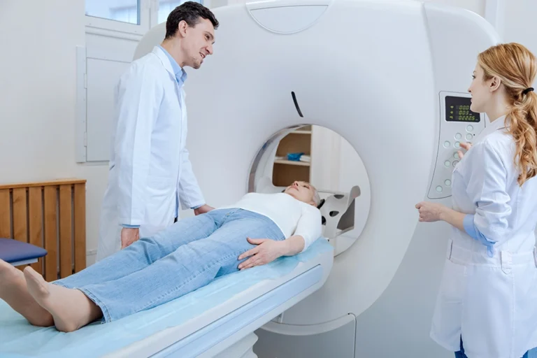
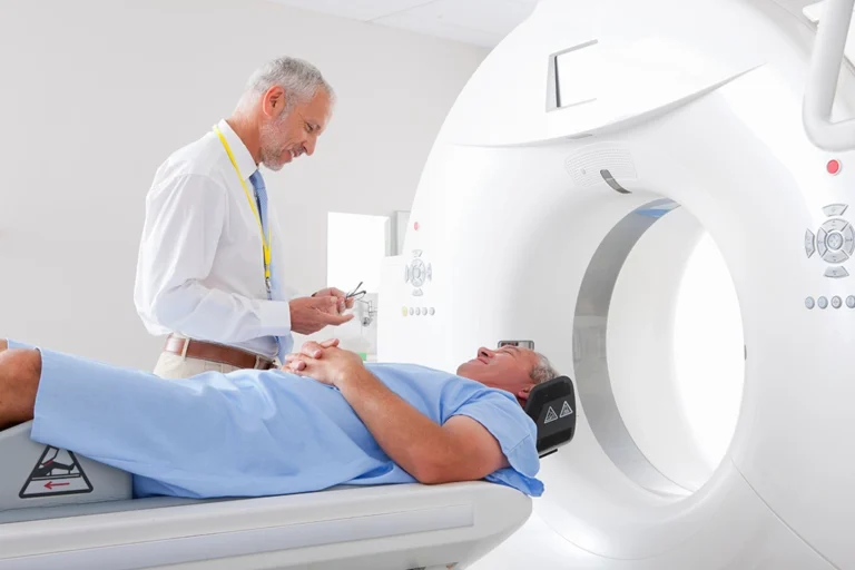
One of the most useful diagnostic tools available in modern medicine today is a CT scan, which medical professionals use to diagnose many different conditions.
CT scans (or computed tomography scans) are used to identify a wide range of diseases and injuries within various parts of the body, like possible tumors within the abdomen or the heart, for example.
CT scans are non-invasive, and are performed relatively quickly. But the best thing about a CT scan is that it can help save your life.
Getting to know the results of your CT scan
Understanding the results of your CT scan can help you be a more active and helpful participant in your health. The better you understand the results of your CT scan, the more you will be able to make good health decisions.
What should I expect when I first look at my CT scan results? How is it organized?
Your CT scan report will be organized into sections, each containing images along with descriptive texts interpreting these images.
If you’re new to CT results, the images from a CT scan may look like just a bunch of images in shades of gray, but once you grasp the main principles of a CT scan, it will become much easier for you to understand.
How are the CT images displayed and labeled in a typical CT report?
The CT images will be arranged in a sequence, each marked and sometimes annotated by a radiologist to highlight important features.
As mentioned before, CT scan images appear in varying degrees of gray, from white to black. These shades measure the density of each of your internal structures, as compared to the next.
Your healthcare team will use something called the Hounsfield Scale, which measures how dense your tissue is, as a guide to understanding your CT images.
Decoding medical terminology in CT results
Medical terminology can seem very complex, especially in a CT scan report. In your report, you’ll also see some anatomical references, descriptions of density and size, and sometimes diagnostic terms.
What are some of the medical terms I should understand in my results?
In addition to the terms already discussed, there are some other terms, such as abnormalities that you should be familiar with:
- Lesion — informal term meaning imaging abnormality, which could be normal or concerning
- Calcification — tissue hardened by calcium
- Cyst — fluid-filled sacs
- Mass — unexpected volume or growth of tissue
- Nodule — lumps of tissue distinct from their surroundings
- Tumor — areas of abnormal cell growth
How should I interpret information about the size or location mentioned in the report?
Size and location descriptions help locate where an abnormality is and how big it is, which is important for diagnosis. Your healthcare provider will interpret any information about the size or location of any abnormality mentioned in the report.
Visual examples of CT scan results
At first glance, the images of a CT scan appear as formations in shades of white, black, and gray formations. With a little time, however, you’ll be able to better understand what the images mean.
What do typical CT images look like?
Typical CT scan images show cross-sectional views of the body and its organs. As we noted above, these views can appear black or white with varying shades of gray that represent different issues. Your healthcare provider can show you exactly what the shapes and colors on your results mean.
What should I look for in my CT images to better understand my health?
When you look at your CT scans, pay close attention to the organs, bones, and tissues. Try to also look for any abnormalities, like tumors, fractures, inflammation, or blockages that may be present.
How are abnormalities usually noted on CT images?
Abnormalities on CT images are usually noted based on differences in density, contrast enhancement, size, shape, location, and border characteristics compared to the surrounding normal tissues.
Radiologists carefully assess these aspects to identify and accurately state abnormalities, and will pass this information along to your provider, who will discuss it all with you.
Next steps after receiving your CT results
Your healthcare provider has the results of your CT scan and they are ready to discuss the results with you. Here are some next steps you should know about.
How should I discuss my results with my healthcare provider?
Ask your healthcare provider to be as open and frank as possible with you about the results of your CT scan. Some of the terminology may be complex so ask them to carefully and fully explain the results to you in layman’s terms that you can easily understand.
When I meet with my provider to discuss my results, what are some good questions to ask?
Here are some questions you should consider asking your healthcare provider about your CT scan results:
If the scan results show any abnormalities, ask your healthcare provider to fully explain these and any implications for your future health.
If there is an issue, ask your healthcare provider about possible future treatment plans and other tests they might order.
How to schedule an appointment with us
Our goal is to offer you and your healthcare provider the most informative results possible, and we make it easy for you to get an appointment.
With numerous locations across South Jersey, you’ll find us conveniently located near major highways and key bridges in the region.
We’ll ensure the entire scheduling process is as effortless as possible for you. Above all, we are here to help you.
Reach out to us at any of the following locations to book an appointment:
- Haddonfield Office – Haddonfield, NJ
- Marlton (Greentree) Office – Marlton, NJ
- Medford Office – Medford, NJ
- Moorestown Office – Moorestown, NJ
- Mount Laurel Office – Mount Laurel, NJ
- Route 73 (Voorhees) Office – Voorhees Township, NJ
- Turnersville Office – Turnersville, NJ
- Voorhees (Carnie Boulevard) Office – Voorhees Township, NJ
- Sewell (Washington Township) Office – Sewell, NJ
- West Deptford Office – West Deptford, NJ
- Willingboro Office – Willingboro, NJ
Learn more about the board-certified, subspecialized radiologists who read, analyze and interpret the findings here at South Jersey Radiology Associates.
Frequently Asked Questions
You will find organized sections, each containing images along with descriptive texts interpreting these images.
The images are arranged in a sequence, each marked and sometimes annotated to highlight important features.
Terms often include anatomical references, descriptors of density and size, and sometimes diagnostic terms.
Size and location descriptions help pinpoint where an abnormality is and how big it is, which is crucial for diagnosis.
Typical CT images show cross-sectional views of the body, which can be in black and white, with varying shades to depict different tissues.
Look for consistency in tissue density and any marked areas that might indicate abnormalities.
Abnormalities may be circled, highlighted, or accompanied by arrows, with annotations explaining the concern.
Discuss any abnormalities, the implications for your health, and any necessary next steps.


